
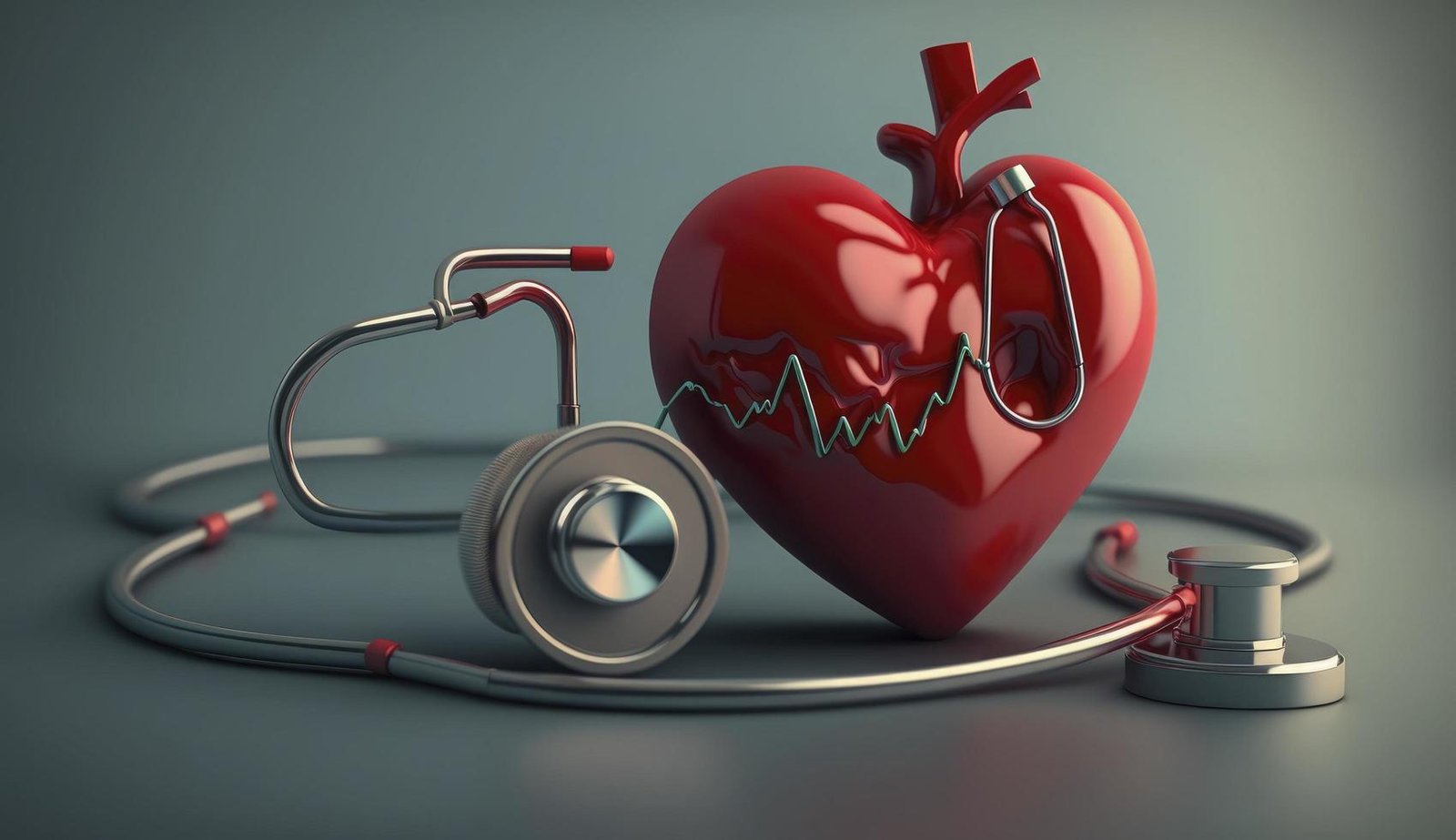

Stethoscope Skills; In the realm of medicine, the humble stethoscope stands as an iconic symbol of the healer’s craft. Yet, its significance transcends mere symbolism; it is a powerful tool in the hands of a skilled practitioner, capable of unlocking vital clues hidden within the body’s symphony of sounds. Welcome to the world of using stethoscopes for differential diagnosis: a realm where each murmur, each rhythm, whispers its own story of health or affliction.
Accorrding to nlm, Originally intended for auscultating chest sounds, stethoscopes serve as vital tools for medical professionals, aiding in the evaluation of cardiovascular and respiratory health. Beyond their primary use, they find application in diverse medical contexts, such as assessing bowel sounds in the gastrointestinal system or detecting vascular bruits.
In this guide, we delve into the art and science of harnessing the stethoscope skills, offering invaluable tips and strategies to sharpen your diagnostic acumen and elevate patient care to new heights. Join us on this journey as we explore the nuances of auscultation, uncovering secrets that lie beneath the surface, and empowering clinicians to make informed decisions that can change lives.
Optimizing Stethoscope Skills
Proper Fit
Comfortable Fit: Adjust the earpieces to fit snugly in your ears without causing discomfort.
Good Ear Seal: Ensure the ear tips create a seal in the ear canal to block out ambient noise and enhance sound clarity.
Diaphragm vs. Bell
Diaphragm Use: Employ the diaphragm for auscultating high-pitched sounds such as lung and heart sounds. Apply firm pressure to enhance contact with the skin.
Bell Use: Utilize the bell for detecting low-pitched sounds, particularly useful for assessing bowel sounds. Apply light pressure to prevent damping of low-frequency vibrations.
Systematic Approach
Chest Examination
Start with the lungs: Listen to breath sounds bilaterally, moving from apex to base and comparing symmetrical areas.
Assess heart sounds: Focus on the four main auscultatory areas (aortic, pulmonic, tricuspid, mitral) using both the diaphragm and bell.
Abdominal Examination
Begin with bowel sounds: Auscultate in all four quadrants, listening for normal, hypoactive, or hyperactive bowel sounds.
Evaluate vascular sounds: Listen for bruits over major arteries, such as the abdominal aorta and renal arteries.
Other Areas (as needed)
Perform a focused examination based on the patient’s complaints or suspected pathology.
For example, assessing for murmurs in the carotid arteries, listening for renal or femoral bruits, or auscultating for abnormal sounds over the thyroid gland.
Documentation
Record findings accurately in the patient’s chart, noting any abnormalities or changes from previous exams. Include the location, intensity, quality, and timing of any abnormal sounds detected.
Maintenance
Regularly clean and disinfect the stethoscope to prevent cross-contamination between patients.
Inspect the tubing, earpieces, and diaphragm/bell for any signs of wear or damage, replacing parts as necessary to maintain optimal performance.
Identifying Key Sounds in Stethoscope Skills
Common Heart Sounds
Normal Heart Sounds (S1 & S2)
S1 (Lub): Occurs with the closure of the atrioventricular valves (mitral and tricuspid valves) at the beginning of systole, signifying the start of ventricular contraction.
S2 (Dub): Results from the closure of the semilunar valves (aortic and pulmonary valves) at the end of systole, indicating the end of ventricular contraction and the beginning of diastole.
Potential Abnormal Heart Sounds
Murmurs: Extra heart sounds caused by turbulent blood flow through the heart valves or great vessels. Murmurs can be systolic or diastolic and may indicate valvular abnormalities or other cardiac conditions.
Gallops: Extra heart sounds resembling the sound of a galloping horse. S3 (Kentucky) and S4 (Tennessee) gallops may indicate abnormal ventricular filling, often associated with heart failure or other cardiac pathology.
Breath Sounds
Normal Breath Sounds
Vesicular: Heard over most of the lung fields, characterized by soft, low-pitched sounds during inspiration and a shorter, softer expiration.
Bronchial: Heard over the trachea and main bronchi, characterized by louder, higher-pitched sounds during expiration than inspiration.
Potential Abnormal Breath Sounds
Crackles (Rales): Discontinuous, popping sounds heard during inspiration or expiration, often indicative of fluid accumulation or airway collapse in the lungs. Can be fine or coarse, indicating different pathologies.
Wheezes: Continuous, musical sounds typically heard during expiration, resulting from narrowed airways due to inflammation, constriction, or obstruction.
Bowel Sounds
Normal Bowel Sounds
Active, Regular Sounds: Characterized by gurgling, clicking, or rumbling noises occurring every 5 to 15 seconds in all four abdominal quadrants.
Potential Abnormal Bowel Sounds
Hyperactive: Increased frequency and loudness of bowel sounds, often associated with diarrhea, gastroenteritis, or early intestinal obstruction.
Hypoactive: Decreased or absent bowel sounds, which may indicate decreased peristalsis due to conditions such as ileus, peritonitis, or bowel obstruction.
Integrating Findings into Differential Diagnosis
Correlating Abnormal Sounds with Potential Causes
Heart Sounds
Murmurs
Systolic murmur at the apex: Mitral valve regurgitation, mitral valve prolapse.
Diastolic murmur at the left sternal border: Aortic valve regurgitation, mitral stenosis.
Gallops
S3 gallop: Left ventricular failure, heart failure with preserved ejection fraction.
S4 gallop: Hypertrophic cardiomyopathy, ischemic heart disease.
Breath Sounds
Crackles (Rales)
Fine crackles at lung bases: Pulmonary fibrosis, heart failure.
Coarse crackles throughout lungs: Bronchiectasis, pneumonia.
Wheezes
High-pitched wheezes: Asthma, chronic obstructive pulmonary disease (COPD).
Low-pitched wheezes: Bronchiolitis, bronchial obstruction.
Bowel Sounds
Hyperactive Bowel Sounds
Epigastric rushes: Gastroenteritis, diarrhea.
High-pitched tinkling sounds: Early intestinal obstruction.
Hypoactive Bowel Sounds
Absent bowel sounds: Ileus, peritonitis, bowel obstruction.
Emphasizing the Stethoscope skills in Comprehensive Evaluation
History
Gather information about the onset, duration, and progression of symptoms, as well as relevant medical history and risk factors.
Physical Examination
Use the stethoscope in conjunction with other examination techniques to assess vital signs, palpate for abnormalities, and observe for clinical signs. Correlate findings from the stethoscope with other physical exam findings to build a comprehensive clinical picture.
Other Investigations
Supplement stethoscope findings with diagnostic tests such as imaging studies (X-rays, CT scans), laboratory tests (blood tests, cultures), and specialized cardiac or pulmonary evaluations (echocardiography, pulmonary function tests). These investigations provide objective data to confirm or refute clinical suspicions derived from stethoscope findings and further refine the differential diagnosis.
Holistic Approach to Differential Diagnosis
Synthesizing Information
Integrate findings from history, physical examination, stethoscope auscultation, and diagnostic tests to develop a prioritized list of potential diagnoses.
Consideration of Context
Factor in patient demographics, comorbidities, and environmental factors to tailor the differential diagnosis and treatment plan to the individual patient.
Iterative Process
Continuously reassess and refine the differential diagnosis based on new information obtained through ongoing evaluation and monitoring.
Tips and Strategies for Enhanced Stethoscope skills
Practice on Healthy Individuals
Auscultate on healthy individuals to familiarize yourself with normal heart, lung, and bowel sounds. Use this opportunity to refine your technique and develop a baseline for recognizing variations in sound quality.
Maintain a Clean Stethoscope
Regularly clean and disinfect your stethoscope to prevent the buildup of debris, oils, and bacteria that can degrade sound quality. Wipe down the earpieces, diaphragm/bell, and tubing with alcohol wipes or a mild soap solution after each use.
Ensure proper storage in a clean, dry case when not in use to prevent contamination.
Consider Electronic Stethoscopes
Electronic stethoscopes offer advantages such as amplification of sounds, noise reduction, and the ability to record findings.Amplification can aid in hearing faint sounds, especially in noisy environments or for patients with obesity or thick chest walls. Recording capabilities allow for playback, analysis, and sharing of auscultation findings with colleagues or for documentation purposes.
Continuously Refine Stethoscopes Skills
Engage in regular practice sessions to maintain and improve your auscultation skills.
Seek feedback from experienced colleagues or mentors to identify areas for improvement.
Utilize online resources, educational materials, or simulation tools to supplement your learning and enhance your proficiency.
Stay Current with Advances in Technology
Stay informed about advancements in stethoscope technology, including new models with improved acoustics or additional features. Attend workshops, conferences, or continuing education sessions focused on stethoscope use and auscultation techniques.
Employ Active Listening Techniques
Focus on actively listening to the sounds you hear, paying attention to nuances in frequency, intensity, and duration. Practice discerning subtle differences between normal and abnormal sounds to enhance diagnostic accuracy.
Integrate Auscultation into Routine Examinations
Make auscultation a routine part of your physical examination for patients across various clinical settings. Incorporate auscultation into bedside teaching sessions to educate patients and trainees on the importance of sound assessment.
Care and Maintenance of a Stethoscope
Caring for and maintaining a stethoscope is essential to ensure its optimal performance and longevity. Here are some key practices for stethoscope care:
Cleaning
Regularly clean your stethoscope to prevent the buildup of dirt, debris, and bacteria. Use a soft cloth dampened with mild soap and water to wipe down the chest piece, tubing, and ear tips. Avoid using harsh chemicals or alcohol-based cleaners, as they can damage the tubing and diaphragm.
Disinfection
After each use, disinfect your stethoscope to eliminate germs and prevent cross-contamination between patients. You can use alcohol wipes or a solution of 70% isopropyl alcohol to disinfect the chest piece and ear tips. Be sure to wipe down all surfaces thoroughly and allow the stethoscope to air dry before storing it.
Storage
Store your stethoscope in a clean, dry place away from direct sunlight and extreme temperatures. Avoid coiling the tubing tightly, as this can cause it to become stiff or kinked over time. Instead, hang the stethoscope around your neck or store it in a protective case to prevent damage.
Maintenance
Inspect your stethoscope regularly for any signs of wear or damage, such as cracks in the tubing or loose ear tips. Replace any worn or damaged parts promptly to maintain optimal acoustics and performance. Lubricate the ear tips and rotating parts of the chest piece occasionally with a small amount of silicone grease to keep them functioning smoothly.
Handling
Handle your stethoscope with care to avoid dropping or mishandling it, which can cause damage to the delicate components. When using the stethoscope, hold it gently and avoid pulling or stretching the tubing excessively. After use, coil the tubing loosely to prevent it from becoming tangled or twisted.
Conclusion
In the symphony of healthcare, mastering the art of auscultation with your Stethoscope Skills is like conducting a precise diagnosis. Just as a skilled musician tunes their instrument, honing your stethoscope skills through practice, cleanliness, and embracing technological advancements ensures you capture every subtle note of a patient’s health.
So, let’s tune in, listen closely, and orchestrate precise diagnoses with every beat and breath. Sound advice: Master your stethoscope skills for a harmonious journey towards better patient care.